The vulva anatomy and internal reproductive organs such as the uterus and ovaries are part of the sexual anatomy commonly referred to as female sexual anatomy. In this article, you will learn about the female genital functions and much more.
Table of contents:
- Labia majora function
- Clitoris function
- The urethra opening
- Female internal genitalia
- Where is the female prostate?
- Female urinary system and where is the female urethra
- Female Urethra Anatomy and Function
- What Is The Mechanism Of Female Reproductive Anatomy?
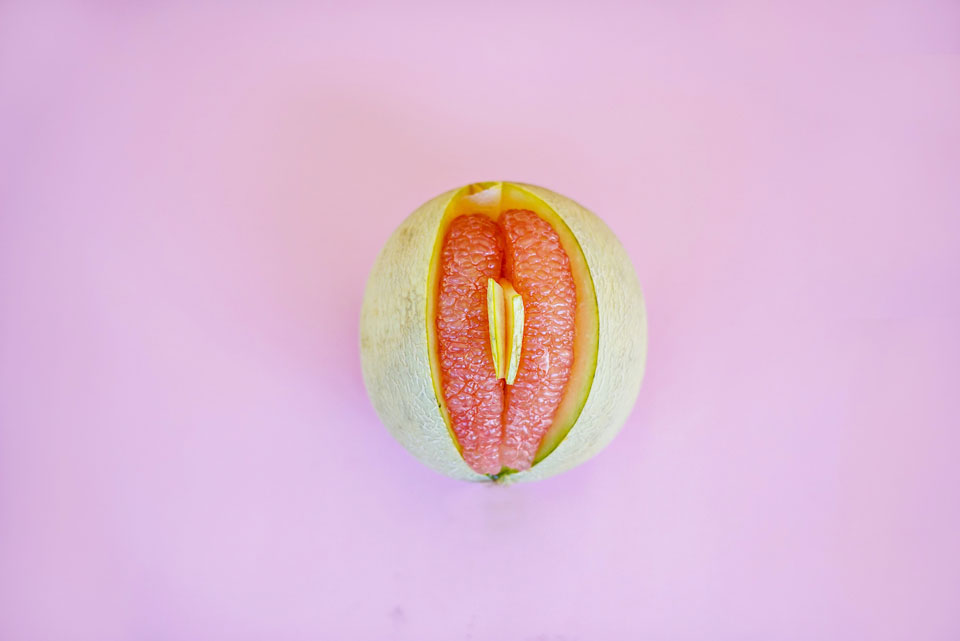
Female external genitalia
Your labia, clitoris, vaginal opening, and urethral opening all belong to the vulva, which is the portion of your genitals that protrude from your body’s exterior.
Many individuals mistakenly refer to the vulva as a “vagina,” even though vaginas are merely one component of the vulva. However, the vulva anatomy and physiology is much more complex than merely the vagina.
Although no two vulvas are identical in appearance, they have the same fundamental components.
Labia majora function
Your vaginal opening is surrounded by skin folds known as labia (lips).The labia majora (external lips) are typically plump and covered in pubic hair.
Your external lips are enclosed by the labia minora (internal lips). They start at the clitoris and finish just below the vaginal entrance.
The labia can be small or large, wrinkly or silky. Frequently, one lip is larger than the next. They range in hue from pink to a dark reddish-black. As you age, your labia’s color may alter. Some people have wider external lips than internal lips, while others have bigger internal lips than external lips.
Both are hypersensitive and enlarge when you are excited. A few sweat glands can be seen on the soft, hairless interior surfaces.
In addition to certain ligaments, smooth muscle fibers, nerves, blood arteries, and lymphatic tissue, the region under the skin comprises fatty tissue.

Clitoris function
Where your internal lips converge at the tip of your vulva is where the head of the clitoris, also known as the glans, resides.
Each person’s varies in size. It can range from the size of a pea to that of a thumb. The clitoral hood encloses the top of the clitoris.
However, this is only the start of the clitoris. Across the two sides of the vagina, it stretches back and down within your body. This component’s stem and crura (roots and legs) measure roughly five inches in length.
The spongy tissue that makes up your clitoris swells when you are aroused. It has more nerve endings than just about any other area of the body, thousands of them.
The urethra opening
The little aperture directly below your clitoris, known as the urethral opening, is where you urinate.
The vaginal opening
Your urethral entrance is just below the vaginal entrance. The vaginal opening is where menstruation blood exits the body, and babies are delivered.
Your vagina may become home to several objects, including hands, male genitalia, sex toys, sanitary pads, and menstruation cups.
Anus
Your rectum’s entrance is called the anus, sometimes the butthole. Because the anus contains so many delicate nerve endings, anal stimulation can be sexually pleasurable for certain people.
Genital area
The fleshy bump over your vulva is called the mons pubis. It protects the pubic bone. It is supposed to be covered in pubic hair after going through puberty.

Female internal genitalia
Female sexual anatomy, or what is often referred to as female, consists of the following internal components:
Vagina
The vulva, cervix, and uterus are all connected by a tube called the vagina. It is the mechanism by which menstrual blood and newborns depart the body.
Some individuals place their sexual partner’s male genitalia, fingers, sex toys, menstruation cups, and/or tampons there. Your genital region is quite elastic and enlarges when you are turned on.
Cervix
Your uterus and vagina are divided by the cervix, which is directly between them. It has a little hole in the center and resembles a doughnut.
This opening joins your vagina and uterus. Sperm can enter, and menstrual blood can exit. During labor, your cervix spreads wide (dilates).
If you stick your middle finger, an erect penis, or a sex object inside your vagina, you can typically feel your cervix near the back of your vagina.
Uterus
The uterus is a muscular organ with a conical shape around the size of a tiny hand. Because a foetus develops there while a woman is pregnant, it is occasionally referred to as the womb.
The bottom portion of your uterus pushes upwards into your lower abdomen during sexual desire. Your vagina lengthens as a result of being turned on. It is known as “tenting”.
Fallopian tubes
Two little tubes make up the fallopian tubes. They deliver eggs to your uterus there from your ovaries. They allow sperm to get through and attempt to fertilise your egg.
Fimbriae
At the tip of every fallopian tube are fimbriae, which resemble little fingers. Every egg is swept into the fallopian tube once your ovary decides to discharge one.
Ovaries
Your eggs are kept in the ovaries. These additionally create hormones such as testosterone, progesterone, and oestrogen. Such hormones regulate processes like pregnancy and menstruation.
The ovaries begin to discharge an egg every month as women approach puberty. The ovaries occasionally discharge several eggs. This continues till menopause.
Glands of Bartholin
Your vaginal entrance is close to where the Bartholin’s glands reside. Once you are turned aroused, they secrete a liquid that moisturises (wets) your vagina.
Skene’s ducts
One on each end of your urethral entrance are the Skene’s glands. They are also known as female prostate glands. They eject fluid when a woman ejaculates.
Hymen
The hymen is a thin, fleshy membrane that spans a portion of the vaginal opening. Hymens differ greatly in the amount of vaginal entrance they span.
Hymens can occasionally (though not necessarily) break and potentially trigger bleeding in the first several instances you insert something into your vagina.
G spot
A few inches within the vulva on the frontal wall is where you’ll find the G spot, also known as the Gräfenberg spot.
When you are turned on, your G spot may enlarge. Sometimes some individuals enjoy getting their G spot rubbed.

Where is the female prostate?
The Skene’s glands are two tiny anatomical structures found only in females. The female genital anatomy says that the prostate is what the glands are.
The urethra and vagina are connected by Skene’s glands, which are situated on each end of the urethra.
The urethra’s entrance is lubricated by the liquid secreted by the Skene glands. The liquid has antibacterial qualities that guard against bacterial infections of the bladder and urinary system.
It’s crucial to remember that these glands discharge into tiny urethral channels. Like the male prostate gland, such glands hold infection far to prevent it from spreading to different body areas.
Understanding the female pelvic physiology is crucial because the Skene glands are located there. Let’s begin, then:
Female anatomy (pelvis)
The pelvis is the bottom portion of the body that lies between the legs and the stomach. In addition to housing the Skene’s glands, the pelvis houses the reproductive system and the intestines.
The female anatomy organs (pelvic) include the following components:
Hip bones
There are two hip bones in the human body, one on each side of the right hip. The pelvic girdle, which includes the hip bones, connects to the top section of the skeleton at the sacrum, where they are attached. Ilium, Pubis, and Ischium are the three minor bones that make up each hip bone.
Sacrum
The lowest portions of the vertebrae are joined by the sacrum. The substantial sacrum bears the weight of the entire body. Five fused vertebrae make up its structure.
Coccyx
The coccyx, sometimes called the tailbone, is joined to the sacrum’s base by several ligaments. It is made up of 4 fused vertebrae that form a triangular structure.
In conclusion, women possess Skene glands, the female equivalent of the prostate. Nearly identical hormones that the prostate glands in males produce are also produced by these glands. Interacting with the pelvis, it plays a significant part in our reproductive systems.
Female urinary system and where is the female urethra
The renal system includes the urethra. This unit contains the bladder, ureters, and kidneys. Urine is the wastewater generated that is produced, stored, and eliminated by the renal system.
Urine deposited in the bladder is transported off the body through the urethra. Since the urethra and reproductive organs are intimately related, males and females have diverse urethral architecture.

Female urethra anatomy and function
The neck, or base of the bladder, is where the female urethra starts. It penetrates the pelvic floor muscles as it descends. Urine flows via the urethral sphincter prior to entering the urethra. This muscle component of the urethra aids in keeping urine within the body before it is expelled.
The vestibule, which is the space among the labia minora, is where the urethra opens. At the border of the vaginal entrance is the urethral opening. The epithelium is a lining that lines the urethra. Mucus is produced by the urethral glands. This mucus aids in preventing acidic urine injury to the epithelium.
Compared to the male urethra, the female urethra is much smaller. As a result, women are more likely than men to get urinary tract infections (UTIs).
What Is The Mechanism Of Female Reproductive Anatomy?
The female reproductive system anatomy serves a number of purposes. The egg cells, also known as ova or oocytes, are produced in the ovaries. The oocytes are subsequently sent to the fallopian tube, where a sperm could fertilise them.
The uterus, in which the uterine lining has grown in reaction to the regular hormones of the breeding process, receives the fertilised egg, and the process continues there. The fertilised egg can lodge into the thicker uterine lining when inside the uterus and proceed to grow there.
In the absence of fertilisation, menstrual flow sheds the uterine lining. The female body‘s reproductive system also creates female sex hormones, which keep the breeding cycle in check.
The female reproductive system progressively ceases producing the female hormones required for the process of reproduction to function throughout menopause. Menstrual periods may now start to fluctuate and finally end. The lady has been regarded as menopausal for one year since her period’s end.
You may also be interested in:
